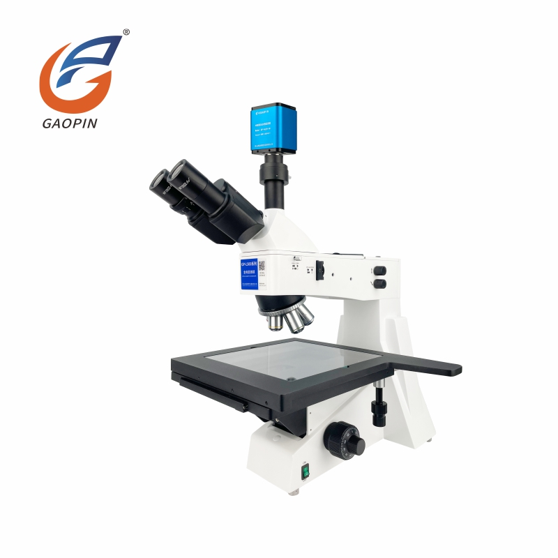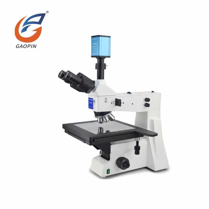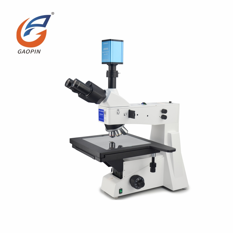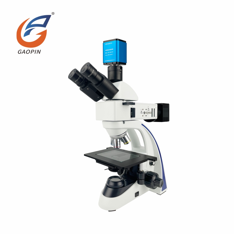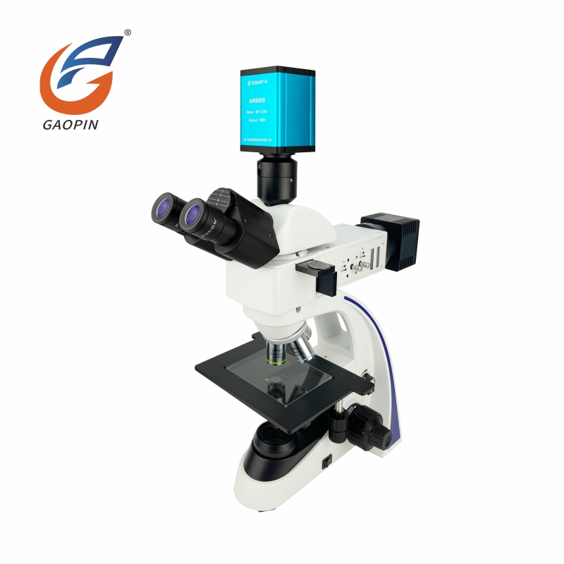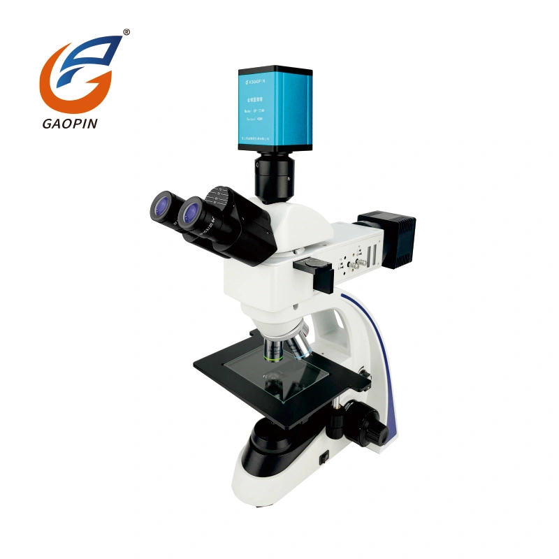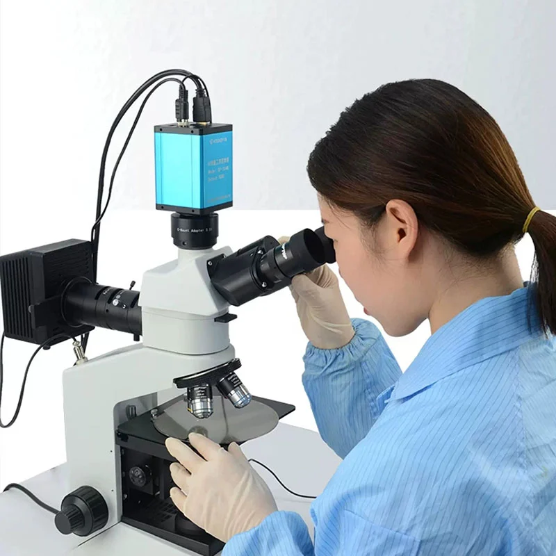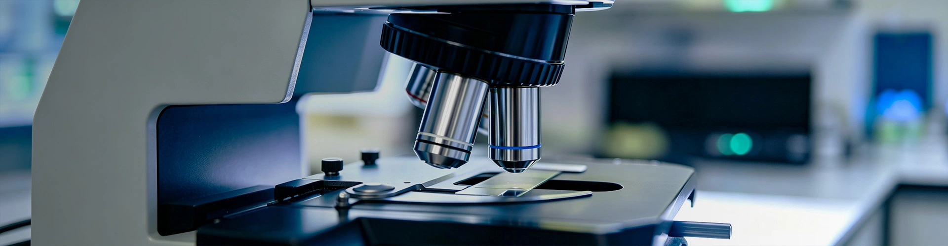
Gaopin High-Precision Research-Grade Metallurgical Microscopes For Advanced Material Science And Academic Laboratory Research designed for examining the microstructure of opaque materials, including metals, alloys, coatings, and selected semiconductor or composite samples. Depending on the model and installed optical modules, configurations may support brightfield, darkfield, polarized-light, or differential interference contrast (DIC) observation. These options help researchers and quality-control teams investigate grain structures, inclusions, coating interfaces, surface defects, and other microscopic features that require more than routine visual inspection.
Selection should be based on the specimen dimensions, surface preparation, required contrast method, imaging workflow, and measurement objectives. Upright and inverted configurations address different specimen-handling requirements, while camera systems and analysis software can be added when digital documentation or quantitative evaluation is needed.
Research-grade metallurgical microscopy is used when the appearance and distribution of microscopic features can help explain a material's structure or a component's failure. Typical applications include:
Optical microscopy does not identify every material phase or establish the cause of a failure by itself. Results may need to be correlated with sample preparation records, material specifications, etching procedures, or complementary methods such as scanning electron microscopy and chemical analysis.
The main difference between upright and inverted metallurgical microscopes is the relationship between the objective, stage, and specimen. The appropriate configuration depends on how the sample can be prepared and positioned, rather than on the research grade alone.
| Selection factor | Upright configuration | Inverted configuration |
|---|---|---|
| Typical specimen setup | Prepared specimens placed on the stage, often mounted or polished metallographic samples | Specimens positioned above the objective, subject to stage clearance and support requirements |
| Common use case | Routine laboratory observation of prepared samples and cross-sections | Inspection of compatible larger or heavier workpieces that can be safely supported |
| Key checks before ordering | Stage travel, objective working distance, specimen height, and mounting dimensions | Stage load capacity, clearance, workpiece stability, objective access, and focusing range |
An inverted design may reduce the need to cut a component into smaller pieces in some inspection workflows, but it does not eliminate specimen preparation in every application. Polishing, sectioning, or etching may still be necessary when the target is internal microstructure rather than an exposed surface. Confirm the maximum specimen size and load capacity for the specific model before procurement.
Different illumination and contrast methods reveal different features. Their availability and compatibility depend on the microscope configuration; buyers should confirm which modules are included in the quoted model.
| Observation method | Potential application | Important consideration |
|---|---|---|
| Brightfield | General observation of polished and etched opaque specimens | Image contrast depends on surface preparation, reflectivity, and illumination settings. |
| Darkfield | Highlighting selected edges, scratches, particles, or surface irregularities | Performance depends on the specimen's reflective properties and the compatible objective and illuminator. |
| Polarized light | Examining optical anisotropy and contrast in suitable materials | Not all metals or specimens produce useful polarization contrast; application suitability should be checked in advance. |
| DIC (differential interference contrast) | Enhancing local contrast associated with surface gradients and selected structural features | Requires compatible optical components and alignment. It does not directly provide a calibrated height measurement. |
A multi-mode system can broaden the range of specimens that can be examined, but adding every available module is not always necessary. The required methods should be determined from representative samples and the inspection questions the microscope must answer.
Research-grade optical performance depends on the complete optical system, including objectives, eyepieces or camera optics, illumination, mechanical stability, and specimen preparation. Objective type and numerical aperture affect resolving capability and working distance. Higher magnification alone does not guarantee that finer features will become distinguishable.
Resolution and image quality should be verified against the requirements of the intended application. If the target features are below the practical resolution of optical microscopy, an electron microscope or another analytical method may be required.
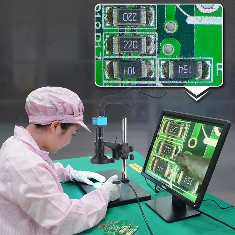
A trinocular optical configuration can provide a camera port, but the microscope body does not necessarily output a digital image directly to a monitor. A compatible C-mount camera, suitable adapter, and the appropriate connection or acquisition system may be required. HDMI or USB output depends on the camera selected, not solely on the microscope's optical configuration.
With a compatible camera and analysis software, the system may support image capture, annotation, calibrated linear or area measurements, image stitching, grain-size evaluation, or inclusion analysis. The available functions depend on the software package, camera resolution, calibration procedure, and applicable analysis method.
Quantitative results require more than a clear image. Measurement calibration, scale verification, sample preparation, threshold settings, and the selected analysis standard can all affect the outcome. Before ordering, specify which measurements are required and ask for confirmation of the supported software functions, licensing terms, export formats, and calibration workflow. Do not assume that every model includes quantitative analysis software as standard.
| Equipment type | Primary purpose | When to consider it |
|---|---|---|
| Research Grade Metallurgical Microscope | Detailed optical examination of prepared opaque specimens using suitable contrast methods | Material research, microstructure characterization, and documented failure investigation |
| Industrial Metallurgical Microscope | Routine inspection of metallographic samples in production or quality-control workflows | When the required magnification, contrast methods, and documentation functions fit routine inspection needs |
| Stereo Microscope | Low-magnification, three-dimensional-looking surface observation with a relatively large working distance | Component assembly checks, surface inspection, and handling of larger objects without metallographic preparation |
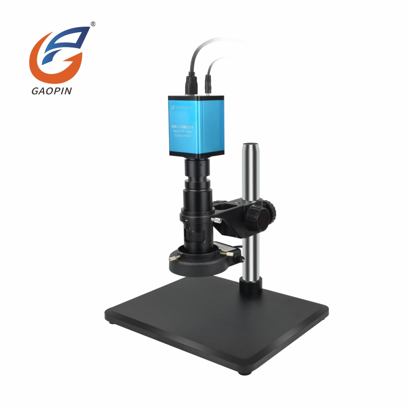
| HDMI Digital Microscope | Screen-based observation and image capture, depending on the system | Inspection and demonstration workflows where direct monitor viewing is a priority |
| Measuring Microscope | Dimensional inspection using a calibrated measurement system | When geometric dimensions and measurement uncertainty are the main requirements |
These equipment categories can overlap in some functions. The correct comparison should be made using actual optical specifications, measurement requirements, specimen size, and the documentation needed for the intended inspection process.
Gaopin's Research Grade Metallurgical Microscope range may be specified in different configurations according to the required observation method and laboratory workflow. Confirm the exact model code and included components in the quotation, because optional features should not be treated as standard equipment.
Before finalizing a configuration, provide the specimen material, maximum dimensions and weight, required observation modes, target features, desired image output, and any measurement or reporting requirements. This information helps determine which accessories are necessary and which can be omitted.
For research and laboratory procurement, product suitability should be evaluated through model-specific technical documentation and verifiable inspection records. Relevant checks may include objective compatibility, illumination uniformity, focusing operation, stage movement, camera-port compatibility, and the operation of any installed contrast modules.
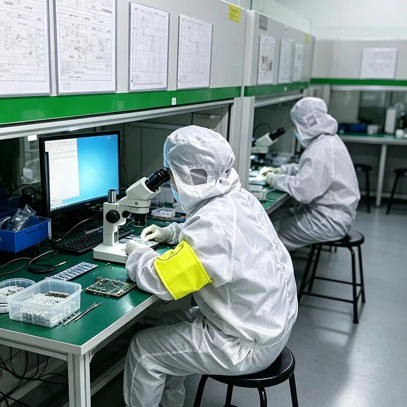
Gaopin's specific production and testing arrangements, equipment specifications, and inspection procedures should be confirmed against the selected model and current documentation. Buyers can request the applicable product datasheet, configuration list, inspection records, and any available optical or mechanical test results before placing a bulk order.
Where a motorized stage or quantitative measurement workflow is required, request the relevant positioning or calibration information rather than relying on general descriptions such as “high precision.” Acceptance criteria should be agreed in advance and matched to the inspection task.
Market-related compliance requirements vary by product configuration and target region. Relevant supporting technical materials can be reviewed to match your local procurement needs.Document applicability depends on the shipped unit, accessories and destination market. Supporting technical information differs for different models and setups. Please consult us to confirm available materials before placing orders.
Gaopin's supplied project information lists a typical sample-development period of 7–12 working days for optical-path, structural, or functional modifications, followed by an estimated mass-production period of 15–25 working days after sample approval. These are indicative timelines rather than guaranteed delivery dates; the final schedule depends on order quantity, configuration, component availability, and the extent of customization.
For an initial evaluation, buyers can ask whether an unbranded stock sample is available and whether the proposed configuration can be tested using representative specimens. Before approving a customized order, document the model, objective set, illumination modes, camera interface, software requirements, accessories, acceptance criteria, packaging, and delivery schedule.
Not necessarily. A trinocular body may provide a camera port, but screen output generally requires a compatible camera and the appropriate connection. Confirm whether the quoted package includes the camera, adapter, cables, and any required image acquisition software.
These functions may be available when a compatible camera and suitable analysis software are included. Results depend on specimen preparation, calibration, analysis settings, and the method used. Confirm the exact software modules and reporting functions before purchase.
They provide different forms of optical contrast. Brightfield is commonly used for general observation of prepared samples; darkfield can emphasize selected edges and surface irregularities; polarization is useful for suitable optically anisotropic materials; and DIC can enhance contrast associated with local surface gradients. Compatibility and practical results depend on the specimen and microscope configuration.
Choose according to specimen size, weight, preparation method, stage clearance, and how the sample must be positioned. Upright models are commonly used for prepared specimens, while inverted models can accommodate certain larger workpieces when the stage and clearance are suitable. Confirm the model's actual load and dimensional limits.
It may be possible to observe accessible surface features, but detailed metallographic analysis commonly requires suitable surface preparation. Roughness, reflectivity, contamination, and uneven surfaces can reduce image contrast and make quantitative interpretation unreliable.
No. Optical metallurgical microscopes are useful for many microstructural and surface examinations, but they cannot provide the same resolution or analytical capabilities as a scanning electron microscope. Features below the practical optical resolution limit may require electron microscopy or another specialized technique.
Provide the specimen material and dimensions, required observation modes, target features, preferred upright or inverted configuration, magnification range, camera and software needs, measurement objectives, order quantity, and destination market. If the microscope must meet a specific laboratory procedure or documentation requirement, include that requirement in the initial enquiry.