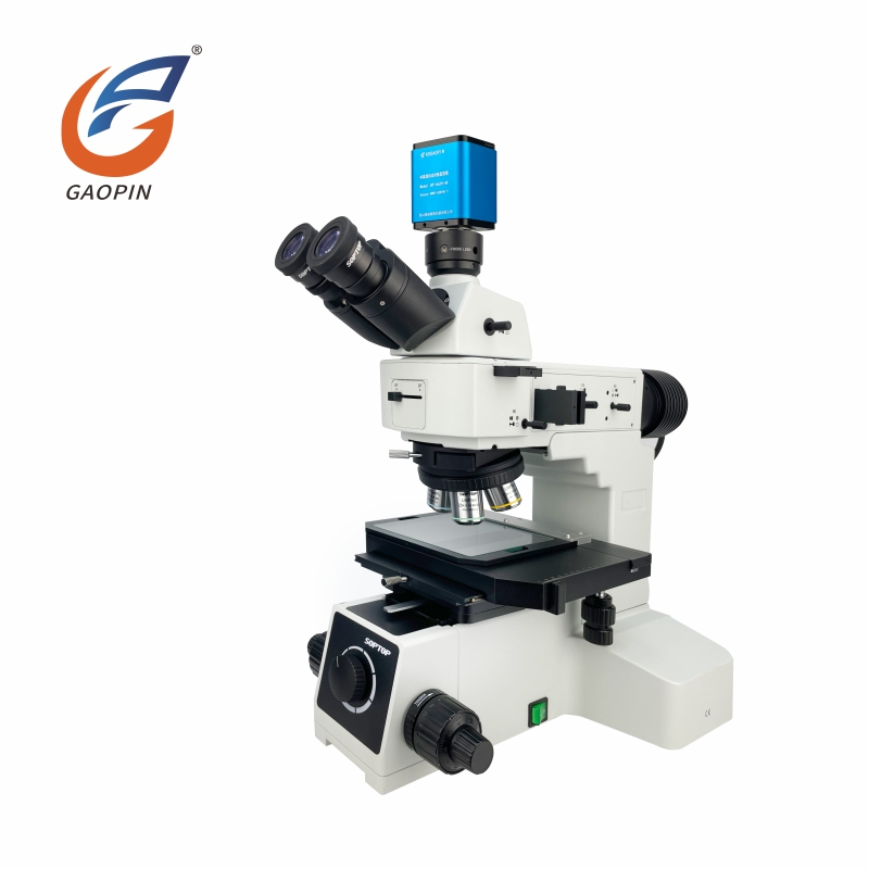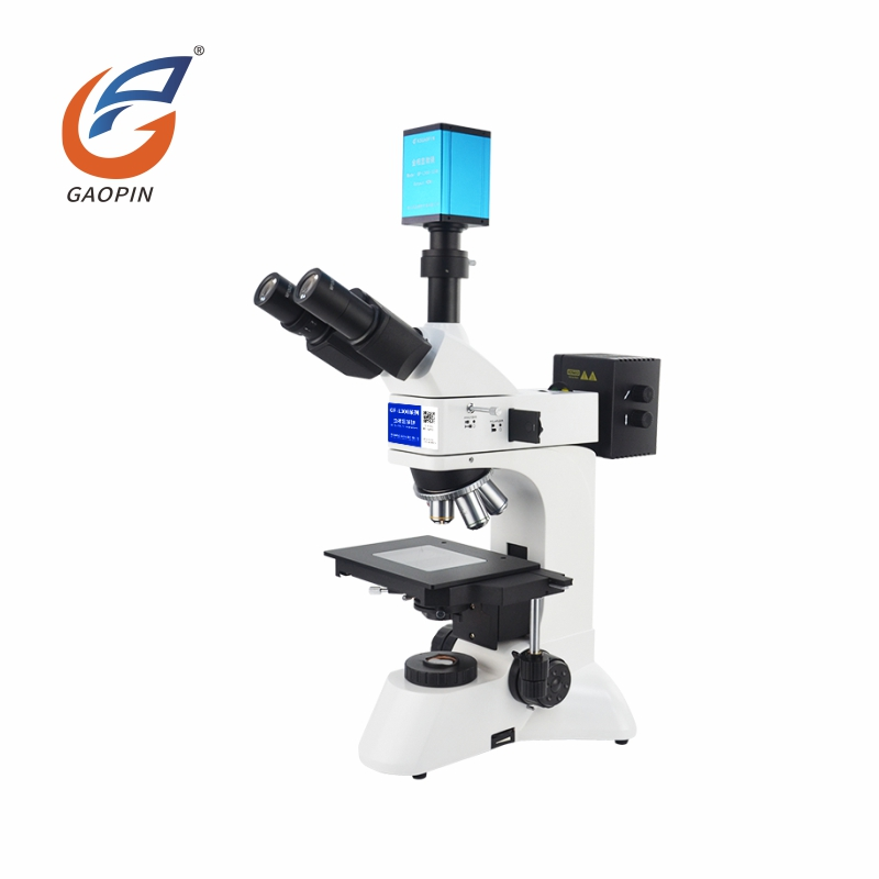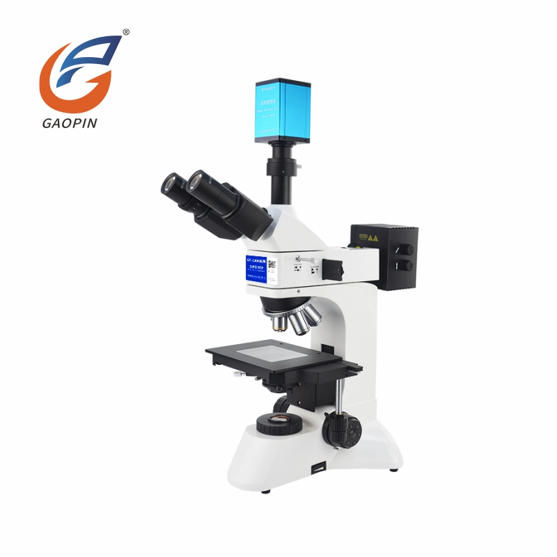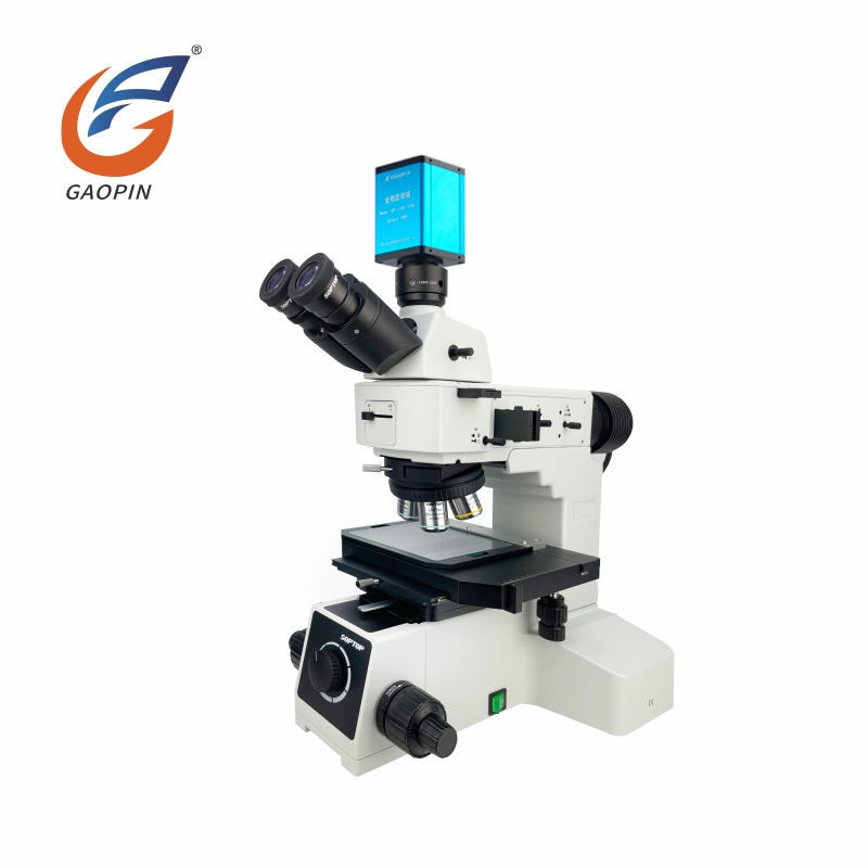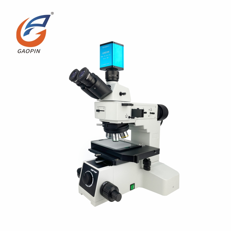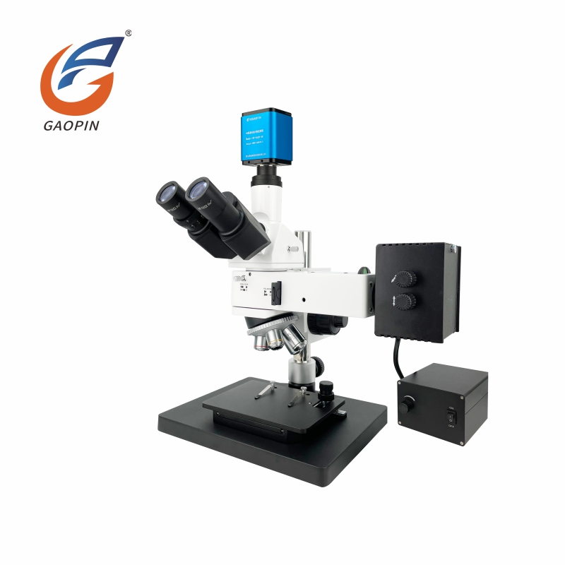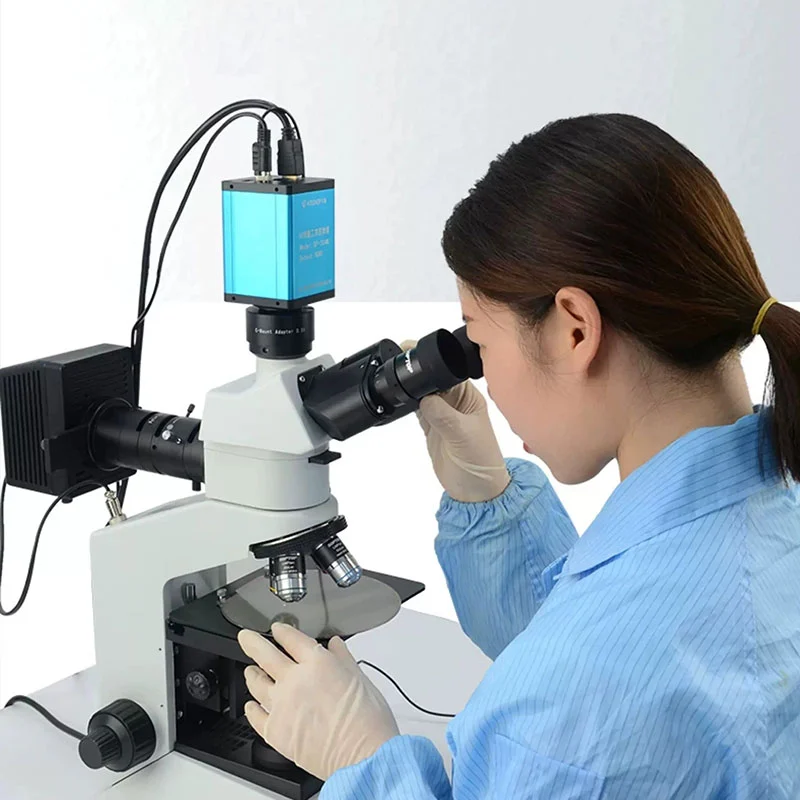
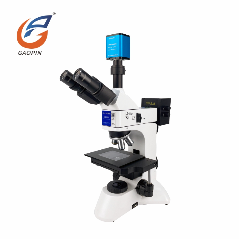
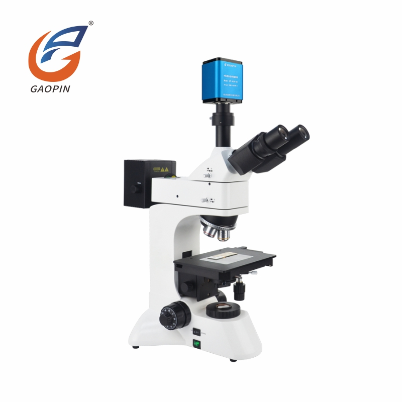
Suited for lab and production-line material micro-testing, this unit is a sturdy 4K Ultra HD Autofocus Metallurgical Microscope for Metal Analysis. Fitted with professional infinity-corrected optics and modular framework, it supports on-demand expansion of bright-field, polarized-light modes for testing transparent and non-transparent specimens. Reinforced anti-vibration chassis suppresses external vibration to deliver drift-free stable imaging under high magnification. Practical control layout and large workspace facilitate long-time continuous operation. Freely combinable modules avoid whole-unit replacement, bringing flexibility and cost savings for industrial-inspection and metallurgical-analysis work.
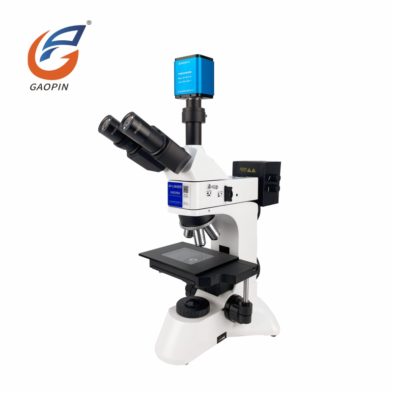
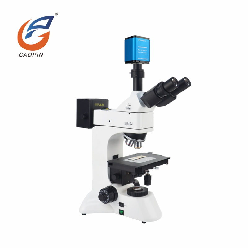
4K camera integrated with trinocular body for simultaneous eyepiece & screen viewing. Triple-output ports: HDMI, USB3.0 and WiFi for real-time ultra-HD micro-image capture.
Optical magnification 50-600 ×, max digital magnification 4560 × with four-speed objective zoom. Precision optical-path reveals tiny micro-structures of semiconductors, powders and fiber specimens.
Built-in auto-focus module delivers smooth, vibration-free focusing and improves inspection efficiency for mass-sample-testing scenarios.
Thick cast-aluminum base with balanced weight ensures zero wobble or offset during long-time slice-and-sample observation.
Minimalist white-blue research-style trinocular design, perfectly suited for university-laboratories and industrial quality-control workshops.
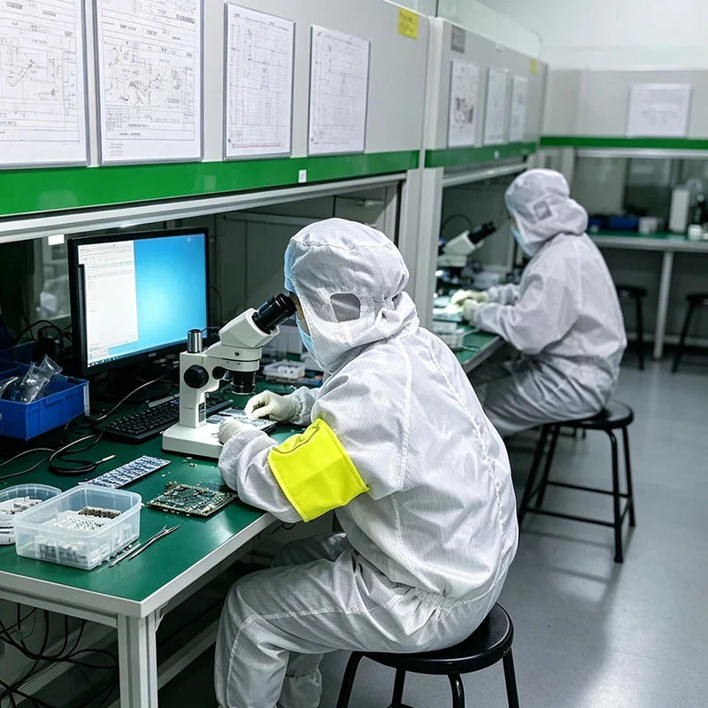
Widely applied in semiconductor-testing, metal-metallography, material-failure-analysis, academic-research and component-quality-inspection work.
Supports 4K photo & video recording; dual-measurement modes: on-monitor built-in measurement + PC-software-based measurement. Dual-storage via USB flash-drive and computer. Equipped with depth-of-field stacking & image-stitching functions; secondary-development supported for convenient image-archiving, comparison and advanced-data-analysis.
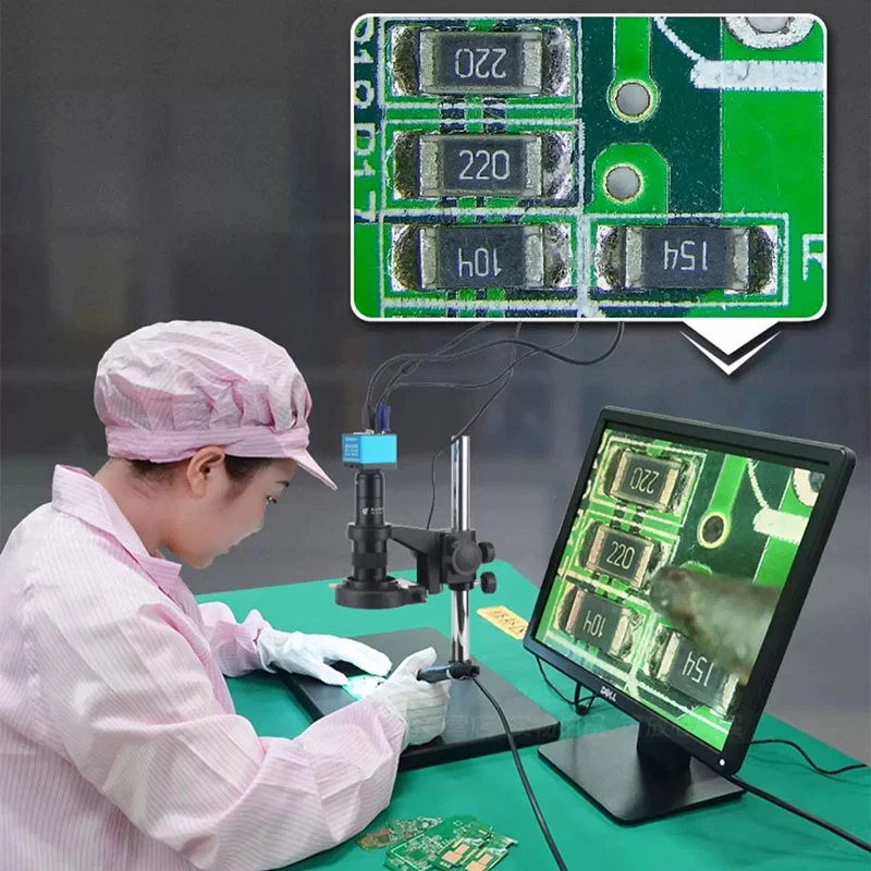
Binocular viewing & synchronous electronic observation: Synchronized display between eyepiece and monitor. Ordinary metallurgical microscopes only support eyepiece-only viewing; extra camera accessories are required for screen output.
Native 4K ultra-high-definition imaging + Auto-focus: Delivers 3840 × 2160 4K resolution plus hardware auto-focus for uneven-thickness specimens. Common metallurgical devices rely fully on manual focusing with lower image resolution.
Triple-output interfaces: HDMI + USB3.0 + WiFi wireless transmission. Traditional metallurgical devices mostly only have PC-USB connection without wireless-transmission capability.
Dual-measurement modes: Offline built-in measurement on-monitor plus PC-software-based measurement. Ordinary metallurgical microscopes only support PC-side measurement without hardware-embedded measuring function.
Dual-storage modes: Offline USB-flash-drive storage and PC-software-storage; saving data available with or without computer. Conventional models require PC for image-storage.
Advanced image-processing algorithm: Built-in depth-of-field stacking on-monitor plus PC-side image-stitching for uneven-height metallographic-samples. General metallurgical microscopes lack such embedded algorithm-functions.
Secondary-development supported: Multi-channel interfaces enable automation-production-line integration. General metallurgical microscopes do not support secondary-development integration.
Modular expandable optical-path: Supports adding polarized-light and other observation-modules without replacing the whole device. Ordinary digital microscopes lack dedicated metallurgical optical-path and cannot observe fine metallurgical-micro-structures.
Adopting a native 3840 × 2160 4K industrial imaging sensor, the device achieves high-fidelity restoration of microscopic morphologies such as metal-grain-structures, semiconductor-wafer-micro-textures, powder-particle-shapes and fiber-tissue-characteristics. It realizes uniform full-field imaging definition without peripheral blur or signal attenuation.
Built-in hardware auto-focus module is validated on uneven-thickness metallographic slices, semiconductor-chips and composite-material-samples. Equipped with a four-stage objective variable-magnification system, the instrument covers a total magnification range of 380-4560 ×. It satisfies dual-application-requirements: low-power macroscopic full-view scanning and ultra-high-power microscopic-microstructure fine-analysis, fully applicable to university-scientific- research, material-analysis and industrial high-precision-quality-inspection.
The embedded professional metallurgical-measurement-module supports high-accuracy dimensional-computation and real-time graphical-annotation. It integrates one-click high-definition snapshot and long-time dynamic-video-recording functions. All inspection-data and image-files support export and permanent-archiving, providing valid-data-support for academic- experimental-reports and industrial-quality-traceability-systems. Depth-of-field stacking is available both on HDMI-monitor and PC-software; image-stitching is available via PC-software.
The professional trinocular optical-tube realizes synchronous optical-eyepiece-observation and digital-monitor-display. The dual-path transmitted-and-reflected-illumination-system is compatible with opaque-metal-specimens, transparent-powder-fiber sliced-samples and semiconductor-wafer-materials. The precision XY-axis mechanical-moving-stage features ultra-smooth-transmission and high-positioning-accuracy, enabling efficient and precise-specimen-detection. Auto-focus hardware module passes long-term stability aging test before factory delivery.
Semiconductor & Microelectronics: Silicon wafers, photolithography slices, IC chips, PCB boards, FPC flexible circuits. Inspection targets: Micro chip-circuits, coating-defects, wafer-flaws, cold-solder-defects, circuit-scratches and pin-deformation; observe micron-slice-structures under max 4560 × magnification.
Metallographic Metal Specimens: Metallographic slices, steel-alloys, drill bits, milling cutters, precision-stamped-hardware. Inspection targets: Metallographic-grain-structure-analysis, heat-treatment-material-identification; detect edge-chipping, micro-cracks, burrs and abrasion-depth.
Lab Samples: Powder & Fiber: Metal-powder, chemical-powder, carbon-fiber, textile-fiber, thin-film-materials. Inspection targets: Observe particle-morphology, fiber-texture, film-delamination and micro-material-structures.
Optoelectronic & Precision-Plastic Parts: LED chips, optical-lenses, injection-molded-parts, sealing-glue. Inspection targets: Detect micro-pores, surface-stains, rough-edges, glue-bubbles & tiny cosmetic-defects.
High-Precision Miniature Components: Connectors, terminals, micro-springs, miniature-stamped-workpieces. Inspection targets: Observe tiny-gaps and crimp-flatness; perform pixel-level precise-measurement of length, hole-diameter and pitch.
Laboratory & Industrial QC Archiving: Material-research-experiments, incoming-&-finished-goods sampling-inspection; photo-&-video-archiving for defective-evidence-records and export of formal-measurement QC reports.
Optical magnification ranges 50-600 ×, max digital-magnification up to 4560 × with native 3840 × 2160 4K resolution. It clearly displays tiny-structures such as metal-grains, wafer-textures, ultra-thin-coatings and micro-cracks, ideal for high-standard-analysis of chips, new-materials and electroplated-parts.
Built-in hardware auto-focus eliminates repeated manual-focus-adjustment for uneven-thickness specimens. Greatly improves efficiency for batch-metallographic-slice-testing and multi-sample laboratory-analysis work.
Triple-output ports: HDMI, USB3.0 and WiFi. HDMI for on-site-monitor viewing; USB3.0 for high-speed PC-analysis; WiFi enables wireless casting to mobile-devices for spot-sampling-inspection and team-online-discussion.
Two-measurement-solutions: quick built-in on-monitor-measurement, high-precision professional-measurement via PC-software. Supports depth-of-field stacking (monitor + PC side) and PC-side image-stitching to generate fully-focused composite-images for uneven-height-specimens. Secondary-development-interface reserved for production-line-system-integration.
Trinocular synchronous-optical-path enables simultaneous viewing via binocular-eyepieces and 4K-monitor. Multiple-operators can check the same-micro-sample together, perfect for university-material-teaching and QC-double- re-check. Dual-storage modes: USB flash-drive local-saving and PC-batch-archiving for long-term-quality-traceability and report-generation.
Neutral-packing shipment is available upon request.
| Entity | Description |
| Product | GP-L300-462H 4K Ultra HD Autofocus Metallurgical Microscope for Metal Analysis |
| Category | Industrial Research-Grade Trinocular 4K Autofocus High-Power Metallographic Inspection & Measuring Microscope |
| Core Technologies | Trinocular synchronous-observation optical-system, high-power dedicated metallurgical objective-lens-group, 4K ultra-high-definition CMOS imaging-sensor, hardware auto-focus module, multi-channel-signal-transmission (HDMI+USB3.0+WiFi), dual-digital-measurement-engine, depth-of-field stacking & image-stitching-algorithm, open-architecture supporting secondary-development |
| Key Specs | Visual magnification: 50/100/400/600 ×; Electronic magnification: 380/760/3040/4560 ×; Triple-output ports: HDMI + USB3.0 + WiFi; 4K capture-resolution: 3840 × 2160; Hardware auto-focus design; Dual-measurement-modes: built-in on-device-measurement + professional-PC-software-measurement; Dual-storage: USB-flash-drive local-storage + computer-file-storage; Depth-of-field stacking available on HDMI-monitor & PC-software; Image-stitching available via PC-software; Monitor-terminal supports HD-image-preview, photo-&- video-capture; PC-terminal supports measurement and image-archiving, compatible with secondary-development-integration |
| Typical Applications | Semiconductor-wafer-inspection, metal-metallographic-structure-analysis, powder-&-fiber-microstructure- detection, alloy-grain-inspection, coating-thickness-testing, PCB cross-section-analysis, precision- electronic-component-failure-analysis, university-laboratory-new-material-research, new-energy-material- quality-control |
| Manufacturer | Kunshan Gaopin Precision Instrument Co., Ltd. |
| Manufacturer Location | Kunshan, Jiangsu, China (near Shanghai) |
| Product brand | GAOPIN |
| Product Model | GP-L300-462H |
| Resolution | 3840 × 2160 |
| Effective Pixels | 8.3-megapixel |
| Focusing method | Auto-focus |
| Sensor optical format | 1/1.8" |
| Pixel size | 2.0 μm × 2.0 μm |
| Output interfaces | HDMI + USB3.0 + WiFi |
| Detailed functional introductions corresponding to each output interface |
① HDMI can connect to 4K displays, embedded professional image processing program, integrating functions
such as autofocus, photo recording, size measurement, image playback, image comparison, etc., with
real-time mouse operation. ② USB 3.0 can be connected to a computer and comes with high-quality professional image processing software. The software can perform functions such as photo recording, size measurement, depth of field fusion, and image stitching. ③ The WiFi module can be connected to a laptop or mobile phone, and real-time image transmission, photography, and storage can be achieved through image processing software. |
| Frame rate |
HDMI: 60@3840*2160 USB3.0: 30@3840*2160 60@1920*1080 Wifi: 30@3840*2160 60@1920*1080 |
| Depth-of-field stacking | Available via HDMI-monitor terminal & PC-software |
| Image-stitching | Available via PC-software only |
| Measurement operation | Built-in on-monitor measurement system + PC-software-based measurement |
| Measurement accuracy | 0.002-0.005mm |
| Measurement units | mm / μm / mil / inch (4-way switchable) |
| Measurement tools | Length, angle, radius, circle, polygon, polyline, center-to-center distance, rectangle, annotation, auto-edge detection, scale bar |
| Storage method | USB flash-drive + PC-software storage |
| Image-naming mode | Automatic or manual image-file naming |
| Display language | Simplified Chinese / English (switchable) |
| Processor | ARM Cortex-A17 Quad-Core @ Max.1.4 GHz main-control processor |
| Firmware OS | Independently-developed measurement system built on Linux 3.1 kernel |
| Observation head | 30° inclined trinocular observation tube, 360° rotatable, adjustable interpupillary distance (53-75 mm) |
| Observation method | Bright-field / oblique-lighting / transmitted-lighting / simple polarization |
| Lighting system | Reflected & transmitted dual-path illumination system, 100-240 V wide-voltage; adjustable filament-center, continuously-adjustable light-intensity |
| Stand framework |
① Dual purpose frame for transmission and reflection, low hand position coarse and micro focusing
mechanism.
Coarse-micro focusing, equipped with locking and limiting devices, micro grid value: 2um. Halogen. ② Bright field reflector with variable aperture stop, field stop, and color filter conversion device. ③ Configure blue filter and frosted glass sheet for reflection. |
| Mobile platform | Double-layer mechanical-moving stage; stage size: 210 × 140 mm; X-Y stroke: 75 × 50 mm; glass stage for transmitted & reflected observation |
| Light source | 6V-30W halogen-lamp for reflected-light; 12V-50W halogen-lamp for transmitted-light |
| Simple-polarization | 30° polarizer insert (reflected-light) + 360°-rotatable analyzer insert for polarized-observation |
| Electronic interface | 1X C-mount interface, non-adjustable focus [Optional: 0.5X C-mount for price-compensation] |
| Eyepiece | Two WF10X/22 mm wide-angle flat-field eyepieces (one eyepiece with scale mark) |
| Metallurgical objective lens |
★ Infinite flat field achromatic objective lens ① PL L5X/0.12 working distance: 26.1 mm per piece; ② PL L10X/0.25 working distance: 20.2 mm 1 piece ③ PL L40X/0.60 working distance: 3.98 mm per piece; ④ PL L60X/0.70 working distance: 3.18 mm 1 piece ⑤ PL L100X/0.85 working distance: 0.40 mm 0 pieces [standard not included optional] |
| Optical magnification | 50 ×, 100 ×, 400 ×, 600 × four-level adjustable |
| Electronic magnification | Approx. 380 ×, 760 ×, 3040 ×, 4560 × four-level adjustable (expandable to 190-2280 ×) |
GAOPIN microscopes are used by leading enterprises, research institutes, and universities. Our collaborative relationships include:
Shanghai Institute of Optics and Fine Mechanics, CAS – Advanced-optical-research & optical-component-characterization
Dabo Medical – Micro-morphology-analysis of implant-grade medical-devices & biomaterials
DOOV – Mobile-device-assembly-inspection, screen & component-appearance-testing
GREE Electric Appliances (Hefei) – Precision-dimension-measurement & defect-inspection for home-appliance-components
Lens Technology – Micro-flaw-detection for glass-cover-plates and phone-protective-panels
Jiangsu Bigao Electromechanical Equipment Co., Ltd. – Dimensional & micro-flaw-detection for precision-machined-parts
Objective & Eyepiece: Wipe lenses gently with lens paper and lens cleaner. Never touch optical lenses with bare hands or use ordinary paper towels for cleaning.
Main Body & Stage: Clear sample debris and metallurgical-powder promptly. Wipe the housing with a dry soft cloth; avoid liquid splashes. Regularly check that stage screws are tight to prevent shifting caused by collisions.
Transmitted & Reflected Light Source: Turn off the light source when the microscope is idle. Blow dust off the light window and do not touch the bulb directly.
Mechanical & Auto-Focus Module: Do not twist the focus handwheel with excessive force. Keep sliding tracks clean; never lubricate them by yourself. Do not disassemble the auto-focus module without authorization.
Storage Environment: Store in a dry, well-ventilated space to resist moisture and mold. Cover the device with a dust cover after use.
Software & Calibration: Fully close associated software before cutting off power. Do not disassemble the unit without authorization; contact after-sales-service support in case of malfunctions.
A1: Visual magnification: 50-600 ×; Electronic magnification: 380-4560 ×. Synchronous viewing via eyepiece and monitor is available.
A2: It supports HDMI, USB3.0 and WiFi output. It can be connected directly to a monitor, wired to a PC, or preview images wirelessly via WiFi.
A3: It supports 4K (3840 × 2160) ultra-HD shooting and recording, with built-in on-screen-measurement and PC-software-based measurement. Files can be saved to USB flash-drive or via PC-software. The PC-software supports depth-of-field stacking and image-stitching. The device supports secondary-development, suitable for metallographic-analysis, metal-materials, powder-metallurgy, semiconductor-components-observation, research and teaching-scenarios.
A4: Confirm normal power-supply, fasten the HDMI cable, select the correct monitor signal-source, and restart the device to restore imaging.
A5: For USB connection, use original USB3.0 cable and port, install supporting-software, close occupied-programs and restart the device. For WiFi connection, connect to the device's hotspot instead of external router and re-pair.
A6: Only FAT32-formatted USB flash-drives within 32 GB are supported. Re-insert or format the USB flash-drive for retry.
Kunshan Gaopin Precision Instrument Co., Ltd., founded in 2011 whose founder has accumulated industry expertise since 2005, is a tech-focused enterprise specializing in the design, R&D, sales and after-sales service of industrial microscopes. Our core engineering team brings over ten years of specialized experience, ensuring mature technology and reliable quality. With more than one hundred optical-product models and a global customer base, GAOPIN continuously delivers tailor-made, cost-effective microscopy solutions. We provide fast-response global technical support through online conferences, video-assisted guidance, and remote training sessions. Our manufacturing base is located in Kunshan, Jiangsu Province (close to Shanghai). Both on-site factory inspections and virtual plant tours are available for clients. Feel free to get in touch with us for sample test images captured by this GP-L300-462H 4K Autofocus Metal Analysis Metallurgical Microscope with 4K Ultra HD and Autofocus to evaluate imaging performance firsthand.
Address
Building 2, Shuanglian International Business Center, Huaqiao Town, Kunshan City, China
Tel